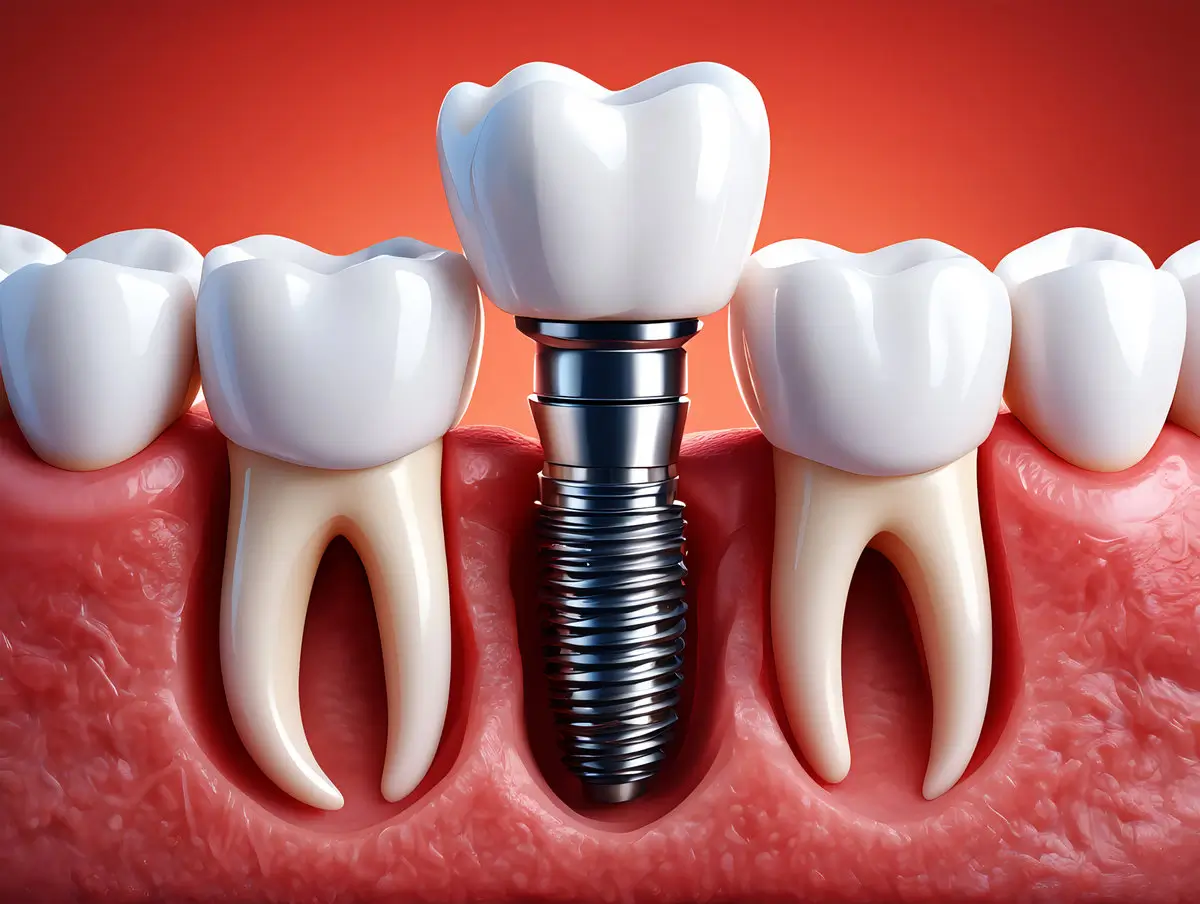
Noticing a change in gum color around your dental implant is understandably alarming, but not every discoloration signals a problem. The color of gum tissue near an implant can range from temporary redness during healing to persistent greyish tones caused by implant materials showing through thin tissue, and each shade points toward a different condition that warrants attention. Understanding what drives these changes helps you decide when to monitor and when to call your oral surgeon.
At Legacy Oral Surgery Group, our team of Dr. Nancy Herbst, Dr. David Farkas, and Dr. Hillel Kaye brings more than 35 years of combined experience to comprehensive dental implant care across our Union City, Elizabeth, and Staten Island locations. We treat more than 7,000 patients annually and monitor gum tissue health throughout the implant process, from placement through long-term follow-up.
Normal Color Changes During Healing
The first weeks after implant placement involve predictable, temporary changes in gum color that reflect your body’s healing response. Fresh surgical sites appear redder than surrounding tissue because increased blood flow to the area is a normal part of tissue repair. This redness fades gradually as inflammation subsides and the soft tissue settles around the implant.
Bruising that creates purple or yellowish discoloration near the surgical site is also common and typically resolves within two to three weeks. As healing progresses, the gum tissue should return to a healthy pink color that blends with your surrounding natural gums. These early color shifts are part of osseointegration, the process by which the implant fuses with the jawbone over the following months.
Grey or Blue Discoloration from Metal Show-Through
One of the most common long-term color changes is a greyish or bluish tint near the gumline, caused by titanium implant components showing through thin gum tissue. Titanium is a grey metal, and when the tissue covering it is naturally thin or translucent, the metal casts a shadow visible from the surface. This is not a sign of implant failure, but it is a cosmetic concern that many patients want to address.
Several factors contribute to metal show-through:
- Thin gingival biotype: Some people naturally have thinner, more delicate gum tissue that masks underlying structures less effectively
- Gradual gum recession: Gums can recede a millimeter or two over time, exposing more of the implant collar or abutment
- Metal abutment material: Traditional titanium abutments are more visible through thin tissue than tooth-colored ceramic alternatives
Two treatments address this reliably. A connective tissue graft, where a small piece of tissue taken from the palate is placed under the existing gum, thickens the tissue and reduces visible grey show-through. Alternatively, switching from a titanium to a zirconia abutment eliminates the grey underlayer entirely. Zirconia is a white ceramic material that reflects light in a way that more closely resembles natural tooth roots, making grey gum color far less likely even in patients with thin tissue.
Signs of Infection: Peri-Implantitis and Peri-Implant Mucositis
Persistent red or purplish discoloration accompanied by swelling, tenderness, or bleeding is a different category of concern. These signs point toward peri-implant mucositis, bacterial inflammation confined to the soft tissue, or peri-implantitis, a more serious infection that involves bone loss around the implant. Research published in the Journal of Clinical Periodontology has found that peri-implantitis affects approximately 22% of implant patients, making it a relatively common complication that requires prompt evaluation.
Inflamed tissue around a dental implant may appear darker or duskier than healthy gum, often with a boggy texture when pressed. Gums that bleed when you brush or floss around the implant, discharge or a persistent bad taste at the site, and a sense that the crown has shifted are all signs that the situation has progressed beyond cosmetic discoloration. Without treatment, infection can silently erode the bone supporting your implant and ultimately lead to implant failure.
Peri-implant disease responds well to treatment when caught early. Depending on severity, options range from professional cleaning of the implant surface and prescription rinses to surgical debridement and bone regeneration procedures. The key is not waiting.
Ready to Schedule Your Consultation?
Our board-certified oral surgeons are here to help you find the right treatment. Schedule your appointment today.
Yellowish or Brownish Staining at the Gumline
A yellowish or brownish tint on gum tissue touching the implant crown sometimes results from surface staining caused by residue from ceramic or composite crown materials, food pigments, or inadequate cleaning around the restoration. This type of discoloration typically appears at the gumline rather than throughout the full thickness of the gum and is more of a hygiene issue than a structural one.
Professional cleaning can usually remove surface staining, though more persistent staining may require polishing or adjustments to the crown margin. Improving your home care routine with a water flosser or interdental brushes designed for use around implants can prevent this type of color change from recurring.
How Implant Placement Affects Gum Color
Some gum color variations are influenced by decisions made during placement and treatment planning. Implant depth relative to the gumline, the angulation of the fixture, and the material and design of the abutment all affect how light reflects through the tissue. When implants are planned with both stability and aesthetics in mind, including a thorough evaluation of tissue thickness and bone anatomy beforehand, the risk of aesthetic color complications decreases significantly.
Front-of-mouth implants in the upper jaw present the greatest cosmetic challenge because they are most visible when you smile. Patients with naturally thin gum tissue in this area benefit from a discussion of abutment material options and tissue augmentation before implant placement, rather than after a grey gumline has already appeared.
Preventing Gum Color Changes Around Your Implant
Good home care dramatically reduces the risk of both infectious and cosmetic gum color changes. Use a soft-bristled toothbrush and implant-compatible floss or a water flosser to clean around the base of the crown thoroughly. Bacterial buildup at the gumline is the primary driver of peri-implant disease, and it is preventable with consistent technique.
Attend professional cleanings on the schedule your oral surgeon recommends, typically every three to six months for implant patients. At these visits, your clinician can identify early tissue changes, measure pocket depths around the implant, and take radiographs to check bone levels. Catching peri-implant mucositis before it progresses to peri-implantitis dramatically reduces the complexity and cost of treatment.
When to Contact Your Oral Surgeon
Color changes that persist longer than three weeks after implant placement, or any new discoloration that appears long after an implant has been in place, should prompt a call to your oral surgeon. Immediate attention is appropriate when discoloration comes with pain, swelling, discharge, bad breath, or any change in how the crown feels during biting.
Monitoring your implant’s long-term success means attending regular follow-up visits even when nothing feels wrong. Most gum color issues respond well to treatment when addressed early, and preventing bone loss around an implant is far more straightforward than repairing it after the fact.
Expert Implant Care at Legacy Oral Surgery Group
Dr. Herbst, Dr. Farkas, and Dr. Kaye evaluate gum tissue health as an integral part of implant monitoring at every visit. Whether a patient is noticing early discoloration after placement or a new color change around an implant that has been in place for years, our team provides thorough examination, accurate diagnosis, and personalized treatment recommendations drawn from decades of combined clinical experience in oral and maxillofacial surgery.
If you have questions about gum color changes around your dental implant or want to schedule an evaluation, we welcome you to contact our office to arrange a consultation at our Union City, Elizabeth, or Staten Island location.
Frequently Asked Questions About Gum Color Changes Around Dental Implants
Is it normal for gums to look grey or darker after a dental implant?
A subtle grey or dark tint can be normal, especially in patients with naturally thin gum tissue. Titanium implant components are grey, and thin tissue allows that color to show through. This is a cosmetic issue rather than a health one, and it can be addressed with a connective tissue graft or a switch to a zirconia abutment. If the color change appears suddenly or comes with pain and swelling, that is a different situation that needs professional evaluation.
What is the difference between peri-implant mucositis and peri-implantitis?
Peri-implant mucositis is bacterial inflammation limited to the soft tissue surrounding an implant. The gums appear red and bleed easily, but bone levels remain stable. Peri-implantitis is a more advanced infection that has progressed to involve bone loss around the implant. Both conditions require treatment, but peri-implantitis is more complex to manage. Early treatment of mucositis can prevent progression to peri-implantitis.
Can grey gums around an implant be fixed without removing the implant?
Yes, in most cases the implant does not need to be removed to correct grey gum discoloration. A connective tissue graft can thicken the gum tissue over the implant, reducing the visibility of the underlying metal. If the abutment is accessible, switching from a titanium abutment to a zirconia one eliminates the grey underlayer. Your oral surgeon can evaluate which option is appropriate based on tissue thickness and the current state of the implant.
How soon after implant placement should gum color return to normal?
Redness and bruising from surgery typically resolve within two to three weeks. If discoloration persists beyond three weeks, or if the color is worsening rather than improving, schedule an evaluation with your oral surgeon. Most post-surgical color changes are normal during active healing, but changes that linger or appear after the initial healing window should be assessed to rule out infection.
What home care steps reduce the risk of gum color changes around implants?
Thorough daily cleaning around the implant base is the most important step. Use a soft-bristled toothbrush and implant-compatible floss or a water flosser to remove bacterial buildup at the gumline. Attend professional cleanings on the schedule your oral surgeon recommends, and do not skip follow-up imaging appointments. Catching early tissue changes before they progress is the most effective way to protect both the gums and the implant itself.