Medically Reviewed by Dr. Nancy Herbst on July 22, 2026
Digital X-rays have replaced traditional film-based imaging at most modern oral surgery practices for good reason. They produce sharper, higher-resolution images with up to 90 percent less radiation than conventional X-rays, display instantly on a computer screen for real-time review, and can be stored, shared, and enhanced in ways film never allowed. For a patient preparing for oral surgery, that combination of safety, speed, and precision makes a meaningful difference.
At Legacy Oral Surgery and Implants in the Elizabeth, NJ area, Dr. Nancy Herbst and our team rely on digital imaging to plan procedures, assess bone structure, and monitor outcomes with confidence. Understanding how digital X-rays work and what they reveal is a useful part of preparing for your care.
What Are Digital X-Rays?
Digital X-rays use advanced electronic sensors instead of traditional film to capture images of your teeth, roots, jaw, and surrounding bone. When the sensor is exposed to a small amount of radiation, it converts that energy into a digital image that appears on a computer screen within seconds. There is no film development, no chemical processing, and no waiting room delay.
The images can be zoomed in, adjusted for contrast, and annotated directly on screen. This allows your surgeon to point out findings in real time and walk you through the diagnostic picture before discussing your treatment options, all in a single appointment.
Types of Digital X-Rays Used in Oral Surgery
Different clinical situations call for different imaging types. Here are the most common:
Bitewing and Periapical X-Rays
Bitewing X-rays capture the upper and lower back teeth on one image and are primarily used to detect cavities between teeth and evaluate bone levels around them. Periapical X-rays show the entire tooth from crown to root tip, along with the surrounding bone, and are particularly useful for identifying infections, abscesses, and bone changes around individual teeth.
Panoramic X-Rays
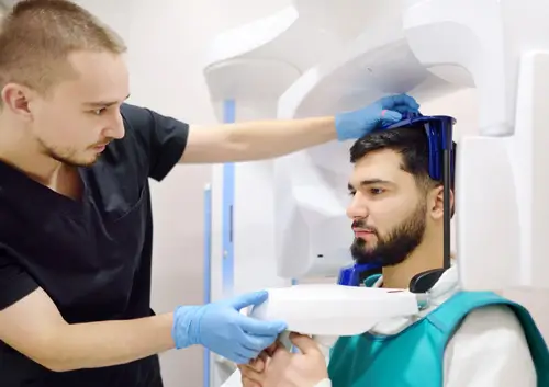
A panoramic X-ray captures a wide view of the entire mouth in a single image, including all teeth, both jaws, the temporomandibular joints, and the surrounding sinuses. This view is valuable for evaluating impacted wisdom teeth, assessing bone quantity before implant placement, and identifying pathology that a series of smaller X-rays might miss.
Cone Beam CT (CBCT) 3D Imaging
Cone beam computed tomography produces three-dimensional images of the teeth, jaws, nerve canals, and sinuses. For procedures like dental implant placement, CBCT imaging is particularly important because it reveals the precise dimensions and density of the available bone and shows the exact location of the inferior alveolar nerve. This level of detail allows for surgical planning that minimizes risk and optimizes implant positioning for long-term stability.
Ready to Schedule Your Consultation?
Our board-certified oral surgeons are here to help you find the right treatment. Schedule your appointment today.
Key Benefits of Digital X-Rays in Oral Surgery
Reduced Radiation Exposure
Research shows that digital X-rays expose patients to 80 to 90 percent less radiation than conventional film X-rays, as documented in peer-reviewed literature including studies published in the National Institutes of Health digital library. For patients who require multiple imaging series over the course of a treatment plan, this reduction is clinically significant. The radiation from a full-mouth digital X-ray series is comparable to background radiation from a short airplane flight.
Immediate Availability and Precision
Images are available within seconds of capture, eliminating the delay of film development and allowing your surgical visit to stay on schedule. The ability to zoom in, enhance contrast, and measure structures directly on screen improves diagnostic accuracy. For oral surgery planning, this precision is not incidental. Identifying the position of impacted teeth, measuring bone volume, or tracing a nerve canal with exactness reduces surgical risk and supports better outcomes.
Easier Storage, Sharing, and Coordination
Digital images are stored electronically and can be shared with referring dentists, specialists, or insurance providers instantly. If you are being treated by multiple providers, your imaging travels with you seamlessly rather than requiring physical copies or re-exposure. This continuity of information can reduce duplicate imaging over the course of a treatment plan.
No Hazardous Waste
Traditional film X-ray processing required chemical developers and fixers that generate regulated hazardous waste. Digital imaging eliminates that entirely, making it both more environmentally responsible and logistically simpler for practices to manage.
How Often Should Dental X-Rays Be Taken?
For patients with healthy teeth and gums and no active oral health concerns, dental X-rays are typically recommended every six to 18 months. Patients with a history of gum disease, frequent cavities, or active oral health issues may need imaging more regularly so that changes can be tracked and addressed promptly. Children and adolescents may require more frequent X-rays during periods of tooth development and eruption.
Your oral surgeon and dentist will recommend an imaging schedule based on your individual history. Because digital X-rays carry minimal radiation exposure, more frequent imaging when clinically warranted carries very little additional risk.
Frequently Asked Questions About Digital X-Rays
Are digital X-rays safe for pregnant patients?
Both the American Dental Association and the American Pregnancy Association consider dental X-rays safe during pregnancy when a lead apron is used to shield the abdomen. The radiation exposure from digital X-rays is very low, and the risk to the fetus is considered minimal. Most dentists and oral surgeons prefer to delay elective imaging until after delivery when possible, but will take X-rays when diagnostically necessary. Let our team know if you are pregnant so we can adjust the imaging plan accordingly.
What is a CBCT scan and when is it used?
A cone beam CT (CBCT) scan produces three-dimensional images of the jaw, teeth, nerves, and sinuses. It is commonly used for dental implant planning because it shows the exact dimensions and density of available bone and the precise location of nerve canals. CBCT imaging is also useful for evaluating complex impactions, diagnosing jaw pathology, and planning corrective jaw surgery. It provides a level of anatomical detail that two-dimensional X-rays cannot match.
How much less radiation do digital X-rays use compared to traditional film?
Digital X-rays use approximately 80 to 90 percent less radiation than traditional film-based X-rays. A complete series of digital X-rays exposes a patient to a radiation dose comparable to the natural background radiation received during a short airplane flight. The minimal exposure makes digital X-rays safe for routine use and appropriate even when more frequent imaging is clinically necessary.
Can I bring X-rays from my general dentist to my oral surgery consultation?
Yes. Recent X-rays from your general dentist are helpful and we encourage you to bring them or have your dentist send them electronically before your appointment. Depending on your procedure, additional imaging may still be needed. For example, if your consultation involves implant placement or a complex extraction, our team may recommend a cone beam CT scan for the three-dimensional detail needed for surgical planning. We will only recommend additional imaging when it is clinically necessary.
Addressing your oral health concerns should not involve guesswork or outdated tools. Our digital imaging technology gives Dr. Nancy Herbst and our team the diagnostic clarity to plan your procedure accurately and explain your findings clearly before any treatment begins. Use our contact form to schedule a consultation and learn how we can help you achieve and maintain a healthy smile.
About the Author
Dr. Nancy Herbst
Dr. Nancy Herbst is a board-certified oral and maxillofacial surgeon with more than 25 years of experience serving patients in Union City, Elizabeth, and Staten Island. She leads Legacy Oral Surgery and Implants, where her practice focuses on dental implants, wisdom tooth removal, bone grafting, and corrective jaw surgery. Dr. Herbst completed advanced training through a hospital-based surgical residency and is committed to delivering precise, patient-centered care using the latest diagnostic technology.